Home > Highlighting JAPAN > Highlighting Japan OCTOBER 2011 > New Diagnostic Method for Creutzfeldt-Jakob Disease
Highlighting JAPAN
SCIENCE
New Diagnostic Method for Creutzfeldt-Jakob Disease
Creutzfeldt-Jakob disease (CJD) is an intractable disease characterized by accumulation of abnormal protein in the brain tissue. A new way to diagnose the disease has now been developed by a team of scientists led by Professor Noriyuki Nishida of the Nagasaki University Graduate School of Biomedical Sciences. The new diagnostic method was published in the medical journal Nature Medicine issued January 30 of this year. Masaki Yamada reports on the latest developments of diagnoses and treatments of CJD, a disease for which most aspects have yet to be revealed.
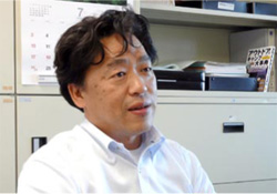
Noriyuki Nishida, professor at the Nagasaki University Graduate School of Biomedical Sciences
Credit: MASAKI YAMADA
Some CJD patients have been confirmed as having eaten meat of cows infected with bovine spongiform encephalopathy (BSE), a transmissible spongiform encephalopathy that is the same type of disease as CJD. Some other patients are confirmed as having received organ transplantations from donors who died of CJD. However, causes of infection remain unknown for about 80% of CJD patients.
CJD occurs in about one of every one million people annually worldwide. Japan has about 130 new cases each year. On average, onset of symptoms occurs at about age sixty, but there are also rare cases of the illness in people in their twenties or thirties.
Initial symptoms of CJD include progressive dementia, which is similar to Alzheimer's dementia, and a variety of other symptoms such as impaired vision, depression and gait disorder. CJD is therefore difficult to diagnose in its early phase. In most cases, CJD infection is confirmed after the illness has progressed considerably or in an autopsy after the patient dies.
"What is extremely important for treatment of CJD," Professor Nishida says, "is to diagnose the disease and confirm the infection at as early a stage as possible."
The only method of antemortem diagnosis of suspected CJD patients used to be brain biopsy, or removal of a small piece of brain tissue for confirming the presence or absence of PrPsc.
However, Professor Nishida notes that the problematic point of brain biopsy is that "removing a piece of brain tissue means damaging a part of the brain. This is highly problematic from the viewpoint of safety. Besides, this method is permitted only for brain surgeons and cannot be performed in hospitals without a brain surgery department."
Clinical studies had revealed that the cerebrospinal fluid of CJD patients has a relatively high concentration of 14-3-3 protein and tau protein*; proteins within nerve cells. This led Professor Nishida to come up with an idea that PrPsc as the cause of CJD may also be detected in the cerebrospinal fluid, which is much easier to take than brain tissue.
Since the conventional detection method does not allow detection of PrPsc, which is extremely small in amount, Professor Nishida developed quaking-induced conversion (QUIC), a new method of in vitro amplification of PrPsc. Professor Nishida's team later made multiple improvements to QUIC and succeeded in developing real-time QUIC (RT-QUIC), a method that permits automatic detection of PrPsc within forty-eight hours.
The team began to apply this method for examining cerebrospinal fluid of suspected CJD patients in Japan (performed by taking the fluid from the lumbar spine via a needle inserted into the back) on consignment from the CJD Surveillance Committee of the Ministry of Health, Labour and Welfare. The team also assessed the technique in a study of about thirty cerebrospinal fluid specimens from patients who died of CJD, which was started in 2009 jointly with an Australian medical team, and achieved greater than 80% sensitivity. On the other hand, the fluid from patients who died of degenerative neurological disorders other than CJD was confirmed as negative with 100% accuracy, which demonstrated the high precision of RT-QUIC. The team is currently conducting joint research with teams from South Korea, Germany, the United States and other countries, in addition to Australia.
"This diagnostic method has enabled us to detect the extremely small amount of PrPsc contained in the cerebrospinal fluid quickly and with extremely high precision," Professor Nishida says, "The method is also safer and easier than brain tissue removal, so diagnosis of suspected CJD patients is now easier than before."
As to whether early diagnosis and early detection will enable treatment of CJD, Professor Nishida says, "unfortunately, there is no established treatment for CJD at the moment. Medicines have been developed that delay the onset of the illness if they are administered in advance, and some of the medicines are being assessed in clinical trials."
Development of new drugs effective for treating CJD is a future issue. Needless to say, however, the achievement of Professor Nishida's team enabling simple diagnosis of CJD by using cerebrospinal fluid is an enormous step forward for the establishment of treatment methods.
*Note: Abnormal tau proteins are considered one of the causes of Alzheimer's disease.


No article or any part there of may be reproduced without the express permission of the Cabinet Office. Copyright inquiries should be made through this form.
© 2009 Cabinet Office, Government of Japan